

Ophthalmology
Neovascular Age-Related Macular Degeneration and Diabetic Macular Edema
Retinal Imaging in Neovascular Age-Related Macular Degeneration and Diabetic Macular Edema: Advancements in Screening and Monitoring


Overview
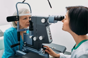
Recent advances in retinal imaging have led to improvements in the early identification and monitoring of patients with neovascular age-related macular degeneration (nAMD), diabetic macular edema (DME), and other retinal disorders. Home optical coherence tomography (OCT) can provide data on the dynamics of retinal fluid exudation and might help to reduce the burden of treatment in the future.
What are some of the important recent advances in retinal screening and monitoring?
Peter Campochiaro, MD
|
|
“Home OCT with AI may allow us to continue to monitor these patients on a long-term basis without the need for frequent office visits.”
I think that one advance in retinal imaging that is particularly interesting is home OCT because it provides opportunities for real-time monitoring and daily retinal assessments, which were previously unavailable. We have become accustomed to assessing patients only when they come into the office, and this can be every 4, 6, or 8 weeks. In between visits, we have little sense of what is going on with the patient. Home OCT gives us the opportunity to obtain that information, and it may be a better way to monitor challenging-to-treat patients who may benefit from more frequent monitoring. Home OCT may also be useful from a research standpoint, as it allows for the real-time monitoring and evaluation of response for a whole group of patients.
Another important advance that is associated with the potential use of home OCT is improvement in the artificial intelligence (AI) software that can assess the amount of exudation that is present and the changes in fluid that occur over time. If changes are noted, the AI software would then send out an alert to the treating physician if there has been a substantial increase in the amount of fluid. The physician can also visit a website that records and monitors the fluid changes.
There is always some apprehension when a new technology is introduced and there are operational logistics to manage. I think that it is important to consider the potential benefits and promise of this technology. Home OCT, for example, will probably not be needed by the majority of patients, but it can be useful in more challenging patients. It is also now being incorporated into clinical trials in which gene therapies, implantable reservoirs, and other long-duration treatments are under investigation.
One of the primary goals of these various treatments is to reduce the number of office visits for patients. In many cases, we are hoping that, with these newer therapies, fewer visits for treatment will be sufficient. Home OCT with AI may allow us to continue to monitor these patients on a long-term basis without the need for frequent office visits.
Peter K. Kaiser, MD
|
|
“It will be very helpful to get patients into the ophthalmologist’s office sooner because we know that an earlier diagnosis leads to better outcomes.”
I think that home OCT is very exciting and is a key innovation to focus on because we are expecting to hear pivotal trial results for one of these technologies shortly. The ability to track treatment response very precisely at home will give us a better understanding of how patients are responding to treatment. One of the obvious benefits of home OCT is that we can identify precisely when a treatment effect appears to be wearing off so that we can act quickly and prevent further deterioration. Home OCT may have a major impact on our current treatment paradigms since many patients have switched from as-needed to treat-and-extend approaches, but we may switch back to an as-needed approach with this technology. Home OCT can hopefully optimize the appropriate treatment interval for individual patients. This is a big step toward personalized medicine, which is going to be a major focus over the next few years.
Currently, screening occurs in retinal diseases primarily through the use of fundus photography to detect diabetic retinopathy. These screening tools can utilize AI to identify patients with certain levels of retinopathy and to identify when they need additional follow-up. However, OCT would be a better technology for detecting patients with macular edema. In the future, the technology that is used in home OCT machines may be able to facilitate the screening and diagnosis of retinal disorders in pharmacies and primary care physicians’ offices.
The home OCT hardware is similar to what we use in our clinics. The resolution that is obtained with home OCT devices is less than what we achieve with an in-office OCT, but the ability to identify potential problems to allow for referral and follow-up with an ophthalmologist is certainly there. The big question: Is the AI performance good enough to make a medical diagnosis in nonoffice settings? AI software has grown by leaps and bounds, just like in other areas of computing. However, the current AI programs are not yet designed for diagnostic applications, although I think that we will see this in the future. It will be very helpful to get patients into the ophthalmologist’s office sooner because we know that an earlier diagnosis leads to better outcomes.
As far as imaging biomarkers that are used in nAMD and DME, there are many biomarkers that are currently being investigated, including subretinal and intraretinal fluid, subretinal hyperreflective material, photoreceptor layer integrity, and others. All of these seem to have some prognostic implications, but, in terms of how we currently use them as clinicians, they have not changed things dramatically because we need additional prospective studies that include these biomarkers to predict outcomes. In the future, these biomarkers could prove to be useful as we continue to move toward personalized medicine to identify patients who may benefit from switching treatments or from more aggressive treatment.
SriniVas R. Sadda, MD, FARVO
|
|
“ . . . we are only beginning to understand the potential utility and benefits of incorporating home OCT into the treatment of patients in clinical practice.”
In the current era of anti-VEGF therapy, we see patients fairly regularly and can identify disease recurrences relatively quickly. With some of the newer treatments that are currently in development, including gene therapies and tyrosine kinase inhibitors that can last anywhere from 6 to 12 months—or potentially even longer—the frequency of patient office visits may decline. Patients receiving some of these newer treatments, however, may eventually require rescue interventions. Without frequent follow-ups, we may miss the opportunity to provide timely rescue in clinical practice if and when it is needed. Home OCT may be an ideal modality for pairing with these longer-acting therapies because it will allow for continued evaluation without additional in-office visits.
I think that it is important to note that most of the clinical studies that have been conducted to date with various therapies that are approved by the US Food and Drug Administration and those in development have been based on fixed patient follow-up schedules and sometimes fixed dosing schedules, which is not necessarily the way that patients are treated in clinical practice. When we follow patients with home OCT, we may begin to see fluctuations in fluid that may be missed with fixed follow-up visits. However, we are still not sure of the amount of additional fluid that should trigger rescue therapy; this has not yet been determined in clinical studies. As we continue to study and gather information on this technology, we are only beginning to understand the potential utility and benefits of incorporating home OCT into the treatment of patients in clinical practice.
References
Fogel-Levin M, Sadda SR, Rosenfeld PJ, et al. Advanced retinal imaging and applications for clinical practice: a consensus review. Surv Ophthalmol. 2022;67(5):1373-1390. doi:10.1016/j.survophthal.2022.02.004
Kumar V, Surve A, Kumawat D, et al. Ultra‑wide field retinal imaging: a wider clinical perspective. Indian J Ophthalmol. 2021;69(4):824-835. doi:10.4103/ijo.IJO_1403_20
Liu Y, Holekamp NM, Heier JS. Prospective, longitudinal study: daily self-imaging with home OCT for neovascular age-related macular degeneration. Ophthalmol Retina. 2022;6(7):575-585. doi:10.1016/j.oret.2022.02.011
Malerbi FK, Mendes G, Barboza N, Morales PH, Montargil R, Andrade RE. Diabetic macular edema screened by handheld smartphone-based retinal camera and artificial intelligence. J Med Syst. 2021;46(1):8. doi:10.1007/s10916-021-01795-8
Metrangolo C, Donati S, Mazzola M, et al. OCT biomarkers in neovascular age-related macular degeneration: a narrative review. J Ophthalmol. 2021;2021:9994098. doi:10.1155/2021/9994098
Oakley JD, Verdooner S, Russakoff DB, et al. Quantitative assessment of automated optical coherence tomography image analysis using a home-based device for self-monitoring neovascular age-related macular degeneration. Retina. 2023;43(3):433-443. doi:10.1097/IAE.0000000000003677
Ong J, Zarnegar A, Corradetti G, Singh SR, Chhablani J. Advances in optical coherence tomography imaging technology and techniques for choroidal and retinal disorders. J Clin Med. 2022;11(17):5139. doi:10.3390/jcm11175139


Explore More in Neovascular Age-Related Macular Degeneration and Diabetic Macular Edema

Neovascular Age-Related Macular Degeneration and Diabetic Macular Edema
Treatment Challenges in Neovascular Age-Related Macular Degeneration and Diabetic Macular Edema
Neovascular Age-Related Macular Degeneration and Diabetic Macular Edema
Exploring the Potential for a Lower Treatment Burden in Neovascular Age-Related Macular Degeneration and Diabetic Macular Edema

Neovascular Age-Related Macular Degeneration and Diabetic Macular Edema
Predicting Outcomes With Anti-VEGF Treatment in Diabetic Macular Edema and Neovascular Age-Related Macular Degeneration

Neovascular Age-Related Macular Degeneration and Diabetic Macular Edema