

Ophthalmology
Neovascular Age-Related Macular Degeneration and Diabetic Macular Edema
The Emerging Role of Artificial Intelligence in Neovascular Age-Related Macular Degeneration and Diabetic Macular Edema


Overview
Artificial intelligence (AI) based on deep learning is playing an increasing role in the detection, diagnosis, grading, and classification of neovascular age-related macular degeneration (nAMD), diabetic macular edema (DME), diabetic retinopathy, and other retinal diseases. Current evidence suggests that deep learning models to detect retinal diseases using multimodal ophthalmic imaging can achieve performance levels that are at least comparable to those that are achieved with operator-based grading techniques and may be an important tool to assist clinicians in personalizing patient care.
Expert Commentary
SriniVas R. Sadda, MD, FARVO
|
|
“ . . . there is excitement about how AI can be used in this era of personalized medicine to identify appropriate treatment approaches and predict outcomes for individual patients.”
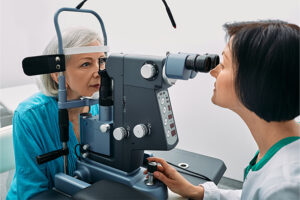
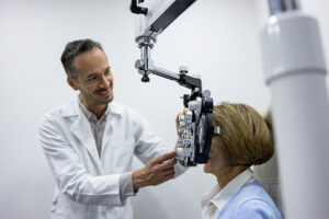
There is significant interest in the use of AI in the diagnosis and management of nAMD and DME. One of the most useful applications of AI in ophthalmology may be in the routine screening for retinal vascular disorders when patients are visiting their primary vision care provider, as well as in the identification of those with abnormal findings who may benefit from a referral to an ophthalmologist or a retina specialist for further diagnosis and management.
A 2018 landmark collaborative study by De Fauw et al evaluating the use of deep learning architecture applied to optical coherence tomography (OCT) scans in patients who were referred to an eye hospital found that the performance in making a referral to an eye specialist for a number of sight-threatening retinal diseases actually reached or exceeded that of the experts. This study highlights the potential capabilities of using AI for screening and referral.
There is also research examining the use of AI in other imaging modalities, such as using color fundus photography in patients with diabetic retinopathy. As more of these screening software tools become available, there will be challenges in garnering regulatory approval and developing successful business models to allow for widespread use.
In terms of other applications, there is excitement about how AI can be used in this era of personalized medicine to identify appropriate treatment approaches and predict outcomes for individual patients. For example, in a patient with nAMD, the use of AI can potentially predict the number of intravitreal injections that they may need given their disease process. AI can also potentially predict how an individual patient will respond to different therapies and how the retina would look after treatment with different therapies.
Researchers in China constructed and evaluated a computer-aided diagnostic system to predict the anti-VEGF therapeutic responses of patients with DME using OCT scans at baseline following the initiation of treatment. A total of 712 patients with DME were classified into poor and good responder groups according to the decrease in central macular thickness after 3 consecutive anti-VEGF injections. Using deep learning algorithms from the OCT scans that were obtained at baseline, machine learning models were constructed to make predictions. The best prediction model achieved a specificity and sensitivity of 0.851 and 0.9, respectively. These values were superior to those achieved by ophthalmologists with several years of clinical experience.
In addition, we have a host of newer treatments on the horizon, such as gene therapy, that are longer acting and target different pathologic mechanisms than many current therapies. Now, how does the ophthalmologist choose the optimal treatment for an individual patient? We have clinical trial data for a population, but, within a trial, individual patients may show a varied response. It is fortunate that we are going to have a convergence between the availability of newer treatments and the availability of improved tools to help clinicians decide which treatment to recommend. All of this feeds into the mantra of personalized medicine, which is providing the right treatment for the right patient at the right time. This is where I see AI helping us going forward.
References
Burlina PM, Joshi N, Pekala M, Pacheco KD, Freund DE, Bressler NM. Automated grading of age-related macular degeneration from color fundus images using deep convolutional neural networks. JAMA Ophthalmol. 2017;135(11):1170-1176. doi:10.1001/jamaophthalmol.2017.3782
Cao J, You K, Jin K, et al. Prediction of response to anti-vascular endothelial growth factor treatment in diabetic macular oedema using an optical coherence tomography-based machine learning method. Acta Ophthalmol. 2021;99(1):e19-e27. doi:10.1111/aos.14514
De Fauw J, Ledsam JR, Romera-Paredes B, et al. Clinically applicable deep learning for diagnosis and referral in retinal disease. Nat Med. 2018;24(9):1342-1350. doi:10.1038/s41591-018-0107-6
Lee CS, Baughman DM, Lee AY. Deep learning is effective for the classification of OCT images of normal versus age-related macular degeneration. Ophthalmol Retina. 2017;1(4):322-327. doi:10.1016/j.oret.2016.12.009
Saleh GA, Batouty NM, Haggag S, et al. The role of medical image modalities and AI in the early detection, diagnosis and grading of retinal diseases: a survey. Bioengineering (Basel). 2022;9(8):366. doi:10.3390/bioengineering9080366
Ting DSW, Cheung CY-L, Lim G, et al. Development and validation of a deep learning system for diabetic retinopathy and related eye diseases using retinal images from multiethnic populations with diabetes. JAMA. 2017;318(22):2211-2223. doi:10.1001/jama.2017.18152

Explore More in Neovascular Age-Related Macular Degeneration and Diabetic Macular Edema

Neovascular Age-Related Macular Degeneration and Diabetic Macular Edema
Treatment Challenges in Neovascular Age-Related Macular Degeneration and Diabetic Macular Edema
Neovascular Age-Related Macular Degeneration and Diabetic Macular Edema
Exploring the Potential for a Lower Treatment Burden in Neovascular Age-Related Macular Degeneration and Diabetic Macular Edema

Neovascular Age-Related Macular Degeneration and Diabetic Macular Edema
Predicting Outcomes With Anti-VEGF Treatment in Diabetic Macular Edema and Neovascular Age-Related Macular Degeneration
Neovascular Age-Related Macular Degeneration and Diabetic Macular Edema